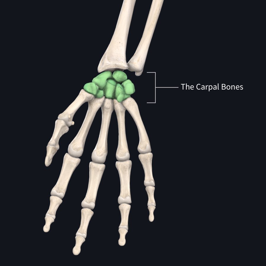
There are 8 small bones that make up the wrist, and this forms a brilliant connection between the forearm and the hand. With this in mind, we are going to take a little journey to discover how the wrist, as simple as it looks, has an important function to stabilize the hand using its surface markings, parts, landmarks, origin and insertion points.
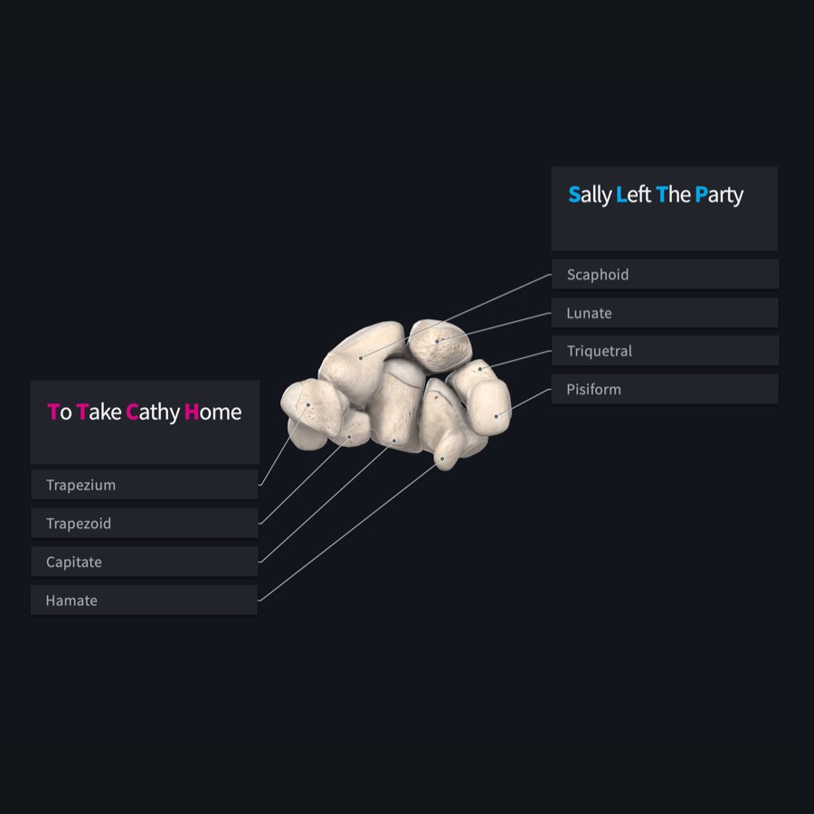
The carpal bones are arranged in two rows. The first row is proximal to the radius and ulna and the second row lies close to the metacarpal bones of the hand. The 8 carpal bones are named thus, from lateral to medial.
Proximal row: Scaphoid, Lunate, Triquetrum, Pisiform
Distal Row: Trapezium, Trapezoid, Capitate and Hamate
“Sally Left The Party To Take Cathy Home”
A popular mnemonic — Sally Left The Party To Take Cathy Home — proves truly useful to remember these special bones.
These 8 bones are named for their unique shapes; Scaphoid (boat), Lunate(Crescent), Triquetrum(3-cornered), Pisiform(pea), Trapezuim(Table), Trapezoid(quadrilateral), Capitate(head shaped), and Hamate (hook-shaped).

These bones are arranged in a special way using their unique and distinct surfaces, landmarks and projections and also with a little help from a special connective tissue known as the Flexor Retinaculum to form a tunnel known as the Carpal Tunnel.
The Carpal Tunnel serves as a channel which transmits all the nerves and arteries from the forearm to the hand, and it also allows tendons from the huge muscles of the forearm to pass through to reach our delicate fingers to allow its movement.
Without these 8 small bones, the hand will have no place to attach, and nerves and blood vessels would have no way to reach the hand. Isn’t it fascinating how all parts of the body work together, and need each other to function optimally?
Discover the most detailed mappings of the human skeletal system ever created in Complete Anatomy 2021. Try it for free today and see for yourself.
If you found this blog post useful, you might also enjoy learning about carpal tunnel syndrome.