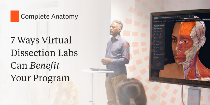
Anatomy Dissected: CNX (vagus nerve)

Cranial Nerve - Vagus Nerve

Welcome back to a brand-new episode of Anatomy Dissected! In this video, we will learn about the tenth cranial nerve, also known as the vagus nerve.  In this episode you will learn:  Check out all the Anatomy Dissected videos on our YouTube channel. READ POST

International Women’s Day 2025

Image of the faces of three diverse Complete Anatomy female models in celebration of International Women’s Day

This year, International Women’s Day is themed around ‘Acceleration Action’. This highlights the need of making moves and clear steps to obtain gender equality. We have made huge strides in this area in the past decade, but it is evident that there is still a lot of work to do. For example, according to an […]READ POST

Highlighting Women’s Health: Complete Anatomy Joins the Rijksmuseum Boerhaave Exhibit

Picture of the Complete Anatomy female model featured in the Rijksmuseum Boerhaave

Photo: Fred Ernst for Rijksmuseum Boerhaave  Shining a Light on Women in Medicine and a Hopeful Perspective for the Future  At Elsevier Health, we continually strive to enhance medical education by reflecting the diverse world we live in. Our latest partnership with the renowned Rijksmuseum Boerhaave in Leiden, Netherlands, highlights this commitment perfectly.  We are […]READ POST

Ganglion Cyst

Image showing the wrist with the skin faded to reveal the bones, muscles, tendons and ganglion cyst beneath.

A cyst is a sac-like structure that is often filled with fluid, or some other substance. There are many types of cysts such as epidermoid cysts, pilar cysts and Baker’s cysts. One type of cyst that is commonly found in the wrist or hand is the ganglion cyst.   Ganglion cysts form near joints or tendons. […]READ POST

Heart Month

Image of the heart with the wall of the right ventricle hidden to reveal the tricuspid and pulmonary valves

This February we celebrate Hearth Month! Heart Month encourages everyone to focus on their cardiovascular health by raising awareness of heart disease and other heart-related conditions. A great way to understand what heart disease is, is to first understand the anatomy of the heart.  The heart is composed of four main chambers. There are two […]READ POST

Diversity and Inclusion in Healthcare

Image of the profile of three dark-skinned models

Historically speaking, anatomical education hasn’t always been diverse. Here at Complete Anatomy, it is well known that we have been aiming to change historical bias when it comes to teaching and learning anatomy. Our first step towards this was introducing our full female model back in 2021 and then our customization options the following year […]READ POST

Anatomy of Glaucoma

Illustration of a dark-skinned female model with the left side of the face showing the underlying anatomy. The lacrimal gland, sclera, cornea, and limbus are labeled

January is Glaucoma Awareness Month. The optic nerve, a bundle of more than 1 million nerve fibers, connects our eyes to our brains, allowing us to see. Glaucoma refers to a group of eye conditions that cause progressive damage to this crucial structure and is a leading cause of vision loss, leaving over 4.5 million […]READ POST

Spermatic Cords

Image of the cross-sectioned testis and spermatic cord.

In the male reproductive system, the testes are suspended by the spermatic cord. Within each spermatic cord are several structures. These include the testicular artery, testicular veins, lymphatics, the deferent duct and testicular nerves.  The testicular arteries arise from the abdominal aorta and descend into the testes. They supply the ureters, testes, and epididymis.   The […]READ POST

Spermatogenesis

Spermatogenesis detailed model showing the process of sperm cell production.

Spermatogenesis is the process whereby spermatozoa are formed. Spermatogenesis has two stages: spermatocytogenesis and spermiogenesis.  Spermatocytogenesis is the first stage of spermatogenesis, whereby spermatogonia develop into spermatocytes and eventually spermatids. Spermatogonia are the germ cells of spermatozoa. Type A dark (Ad) spermatogonia undergo mitotic division at irregular intervals giving rise to either a pair of […]READ POST

Male Pelvis: How it started vs. How it’s going

Side by side comparison of posterior view of the bladder, prostate, seminal glands, and ampulla of vas deferens. Left has the previous model and right has the new model.

Have you checked out Complete Anatomy’s new male pelvis yet? Available now is a brand new, fully remodeled and improved male pelvis!   To highlight its improved detail and accuracy, we have pulled together some side-by-side comparisons. With the help of subject matter experts, 3D artists and our medical team you can now see the effort […]READ POST