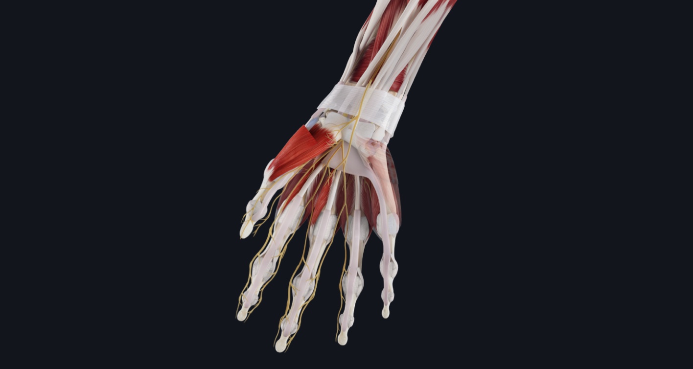
Did you know that the carpal tunnel is a passageway in your wrist? This passageway exists between the tubercles of the scaphoid and trapezoid bones on the lateral side as well as the pisiform and the hook of the hamate on the medial side. The carpal tunnel is deep to the flexor retinaculum and the median nerve travels through it to enter the hand ✋.
The median nerve supplies the 1st and 2nd lumbricals alongside 2 and a half thenar muscles. The median nerve also provides sensory innervation to the entire palmar surface and parts of the digits. It is important to note that the palmar cutaneous branch of the median nerve, which supplies the central palm, does not pass through the carpal tunnel and passes superficial to it instead.
Carpal tunnel syndrome is caused by compression of the median nerve as it travels through the carpal tunnel of the wrist. This causes the myelin sheath and axon to develop lesions while the supporting connective tissues suffer from inflammation. It is the most widespread nerve dysfunction, representing 9 out of every 10 neuropathies. Like other neuropathies, the symptoms of carpal tunnel syndrome include pain, numbness, and tingling of the skin. In carpal tunnel syndrome these symptoms begin in the thumb, index finger, middle finger, and thumb side of the ring finger but may travel up the affected arm over time 💪.
To learn more about the structure of the median nerve and other nerves of the peripheral nervous system, you can check out the peripheral nerve fiber microanatomy model in Complete Anatomy.
If you found this blog post useful, you might also enjoy learning about a crafty mnemonic for the carpal bones.