Pictured above is an exclusive preview of our new Scalp & Meninges detailed model, coming this summer to Complete Anatomy.
The brain is often considered the most protected organ in the body. Let’s look at some of the structures which surround it, starting from the most superficial layer, the scalp.
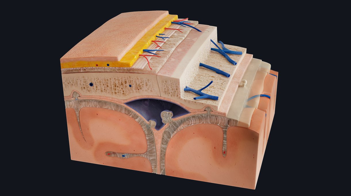
5 layers of tissue make up the scalp, easily remembered by the use of a mnemonic: SCALP.
S – Skin
Just like the skin covering the rest of the body, it acts as a barrier to the external environment. It also has endocrine functions including the formation and synthesis of vitamin D.
C – Connective tissue
The connective tissue of the scalp is known as superficial fascia. It is the most vascularised part of the scalp and functions to connect the skin of the scalp to the layer beneath known as the aponeurosis.
A – Aponeurosis
The epicranial aponeurosis is loosely connected to the underlying tissue and can move freely over it, carrying with it the skin and superficial fascia. It also serves as the site of origin, and a site of insertion for muscles which make up the occipitofrontalis.
These top 3 layers of the scalp are often referred to as the scalp proper.
L – Loose connective tissue
This layer allows for movement of the scalp proper over the calvaria (skullcap).
P – Pericranium
This layer is also known as the periosteum of the skull. It provides structural support as well as a small amount of blood supply to the cranial bone which it adheres to.
Separating the scalp and meninges are the cranial bones. There are 8 cranial bones, held together by joints called sutures.
The meninges are found beneath the cranial bones. They consist of three membranes which surround and protect the central nervous system by anchoring it to the surrounding bones and also produce shock absorbing cerebrospinal fluid.
They are:
Cranial dura mater
A thick, tough and durable membrane. The dura mater is made up of 2 dural layers, the periosteal and meningeal layer.
Cranial arachnoid mater
Arachnoid stems from Greek, meaning spider-like. This mater was named so based on its characteristic web like appearance formed by its wispy fibres.
Cranial pia mater
A delicate vascular membrane, the pia mater attaches directly to the surface of the brain. It translates as “tender mother”, indicating its role in protecting the brain carefully.
A crafty way of remembering the meninges of the brain is by using the mnemonic PAD; Pia, Arachnoid, Dura. Remember the meninges pad the brain, protecting it.
See why Complete Anatomy is head and shoulders above the rest, with the world’s most advanced 3D anatomy platform, including a vast range of detailed models. Try it for FREE today.