Lying within our abdomen is the pancreas. This organ is an accessory organ of the digestive system and has both endocrine and exocrine functions that help us digest the food we eat.
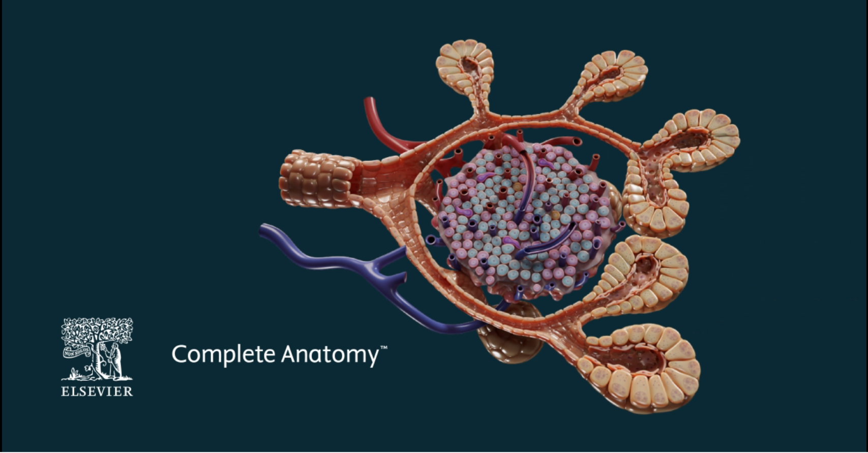
Pancreatic exocrine cells, or acinar cells, are surrounded by loose connective tissue to form clusters of lobular acini. This exocrine unit makes up around 98% of pancreatic tissue. Each acinus is drained by a single intralobular duct. Functionally, the acinar cells secrete zymogens which are inactive forms of enzymes that digest proteins, fats, and carbohydrates. They also secrete water and bicarbonate. These secretions are directed into the intralobular ducts which flow to larger ducts until they reach the pancreatic duct and descend to the duodenum. Enterochromaffin cells located in the acini also secrete several hormones.
The endocrine islets (of Langerhans) make up about 1-2% of the pancreatic tissue. These clusters of cells produce hormones that are secreted directly into the dense capillary beds that permeate the islets. Within the islets, alpha, beta, delta, epsilon and F cells sit. The different cell types in the islet dictate endocrine function. Beta cells secrete insulin which helps decrease blood glucose levels. Alpha cells secrete glucagon which raises blood glucose levels. Delta cells secrete somatostatin which reduces secretion of insulin and glucagon. Epsilon cells secrete ghrelin which is associated with feelings of hunger. F cells help regulate gastrointestinal secretions and motility.
All these features and more can be seen in our new pancreas detailed model in Complete Anatomy, be sure to check it out along with four new detailed models!