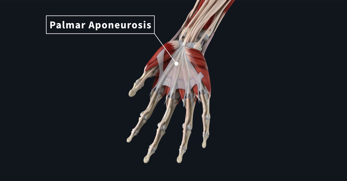
Beneath the skin on the palm of your hand lies the palmar aponeurosis. In the central part of the deep fascia, this thickened structure is highly specialized. It is in the shape of a triangle and protects underlying neurovasculature and tendinous structures. With its firm attachment to the palmar skin, it provides an improvement on grip.
Some structures that lie beneath its protection are:
- the superficial palmar arch
- long flexor tendons
- the terminal part of the median nerve
- the superficial branch of the ulnar nerve
The palmar aponeurosis has several attachments. The apex lies proximal to the flexor retinaculum where it blends with its fibers. It is continuous with the tendon of palmaris longus. The base starts at the distal border of the flexor retinaculum and fans out distally to each base of a finger.
You can divide the palmar aponeurosis into two parts. A superficial strata attached to the dermis, and a deep strata. The deep strata divides into four slips for each finger. These slips divide into two parts that are continuous with the fibrous flexor sheaths. The extensions of the slips pass the deep transverse metacarpal ligament, capsule of the metacarpophalangeal joints and the sides of the bottom proximal phalanx. When you extend your fingers, soft tissue on the palm can be seen bulging out through the four slips.
Dupuytren’s contracture results from a thickening, shortening and inflammation of the ulnar side of the aponeurosis. Resulting in the proximal and distal fingers becoming flexed or present with nodules that form contracture deformities. Treatment options include needle aponeurotomy, collagenase injection, or surgical resection and fasciectomy.