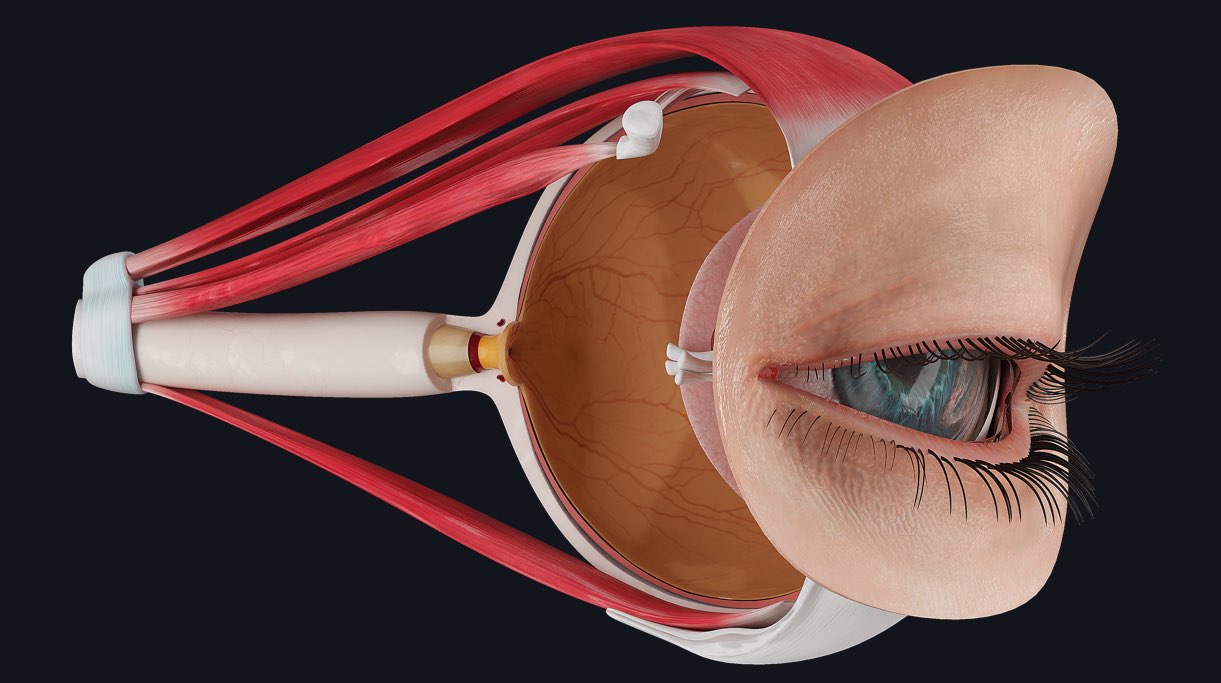
Funduscopy is part of an assessment of the eye performed by a clinician. It is done by inspecting the fundus of the eyeball ?, with an instrument known as an ophthalmoscope (funduscope), which is simply a light source modified optically with the addition of lenses. ?
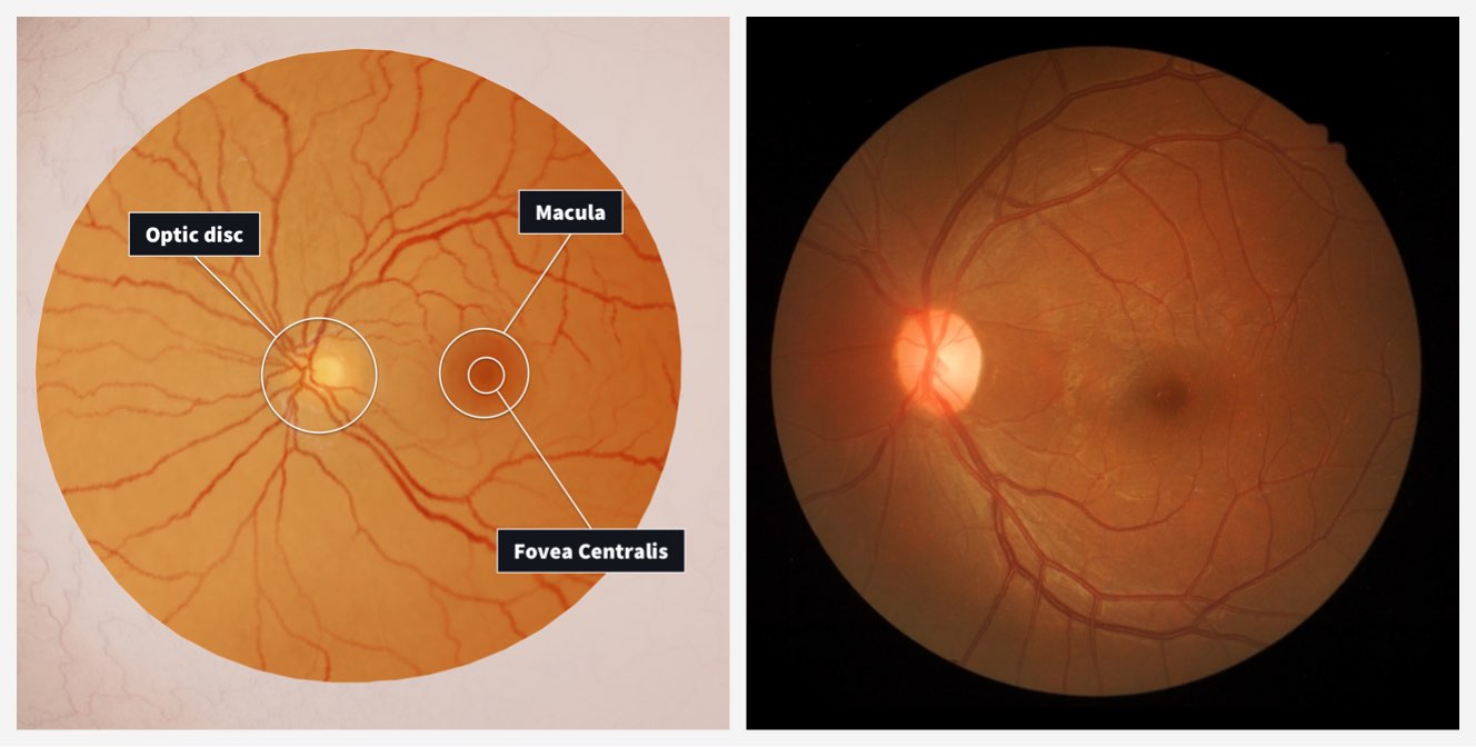
The fundus of the eyeball (pictured above in 3D) is the name given to the posterior aspect of the eyeball that light entering the eye focuses on. Before a funduscopic examination is performed a sound knowledge of the anatomy of the fundus is vital. Let’s look at some distinctive features of the retina in this region:
- The retina contains a circular area called the optic disc where sensory nerve fibres and blood vessels travelling in the optic nerve enter the eyeball. This part of the eyeball is insensitive to light, and it is known as the ‘blind spot.’
- Lateral to the optic disc is a small oval area called the macula, or central retina. It contains specialized photoreceptors known as cone cells which are responsible for the sharpest of vision.
- At the centre of the macula there is a depression called the fovea centralis. This region has the highest density of narrow and elongated cone receptors to maximise light detection.
Before the eye is examined, a thorough clinical history is required to exclude or include differential diagnoses. The pupil is usually dilated pharmacologically for a complete examination of the fundus. ?
During the examination, the clinician usually makes an assessment of the shape, colour, edges and cup size of the optic disc; follows each blood vessel checking for any abnormalities, and finally looks at the macula for any forms of degeneration.
When last did you get your eyes tested and why?
The eye prosection is one of 15 detailed models in Complete Anatomy. Experience the learning power of the most advanced 3D anatomy models available. See for yourself with a FREE 3-day trial.