Complete Anatomy

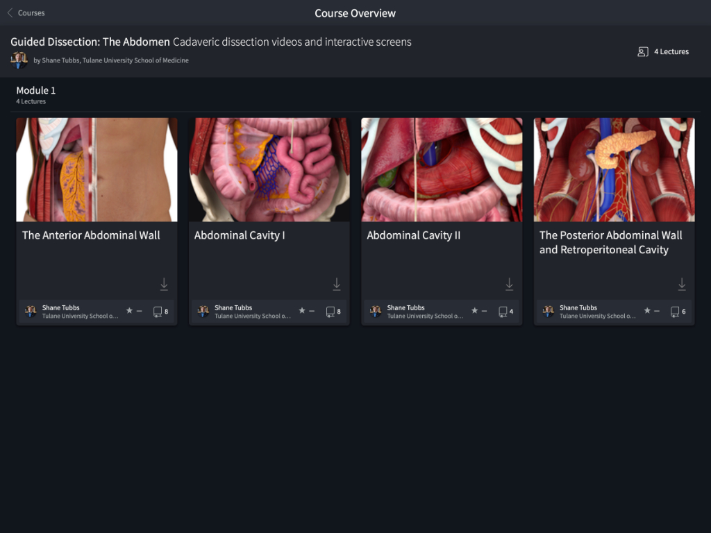
Guided Dissection: The Abdomen

play_circle_filled WATCH THE VIDEO

course poster

About this course

By Dr. Shane Tubbs

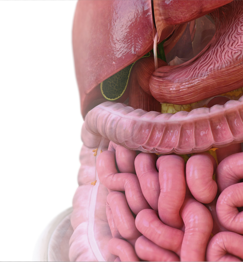
Experience our first virtual dissection built in collaboration with the Gray’s Anatomy family of products. Follow an abdominal dissection from the superficial aspects of the anterior abdominal wall, the major organs and their blood supply, right through to the retroperitoneal space. Combining Complete Anatomy’s detailed virtual models and dissection videos from ‘Gray’s Surgical Atlas,’ this course will provide you with a clear and systematic guide of the abdominal region.

screenshot
screenshot
screenshot
screenshot

Leverage gold standard dissection content by Dr Shane Tubbs, from Gray’s Surgical Atlas.

Follow dissections through the entire abdomen, from the anterior abdominal wall, the abdominal cavity, to the retroperitoneal space.

Correlate video content to Complete Anatomy’s updated abdominal region in real time.

Also available in three languages, Chinese, Spanish, and English.

0LECTURES
0SCREENS
0VIDEOS

Learning Outcomes

done

Identify the layers which make up the anterior abdominal wall.

done

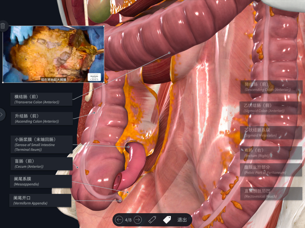
Identify the position of major organs within the abdominal region.

done

Understand the main blood supply and drainage to the foregut, midgut, and hindgut.

done

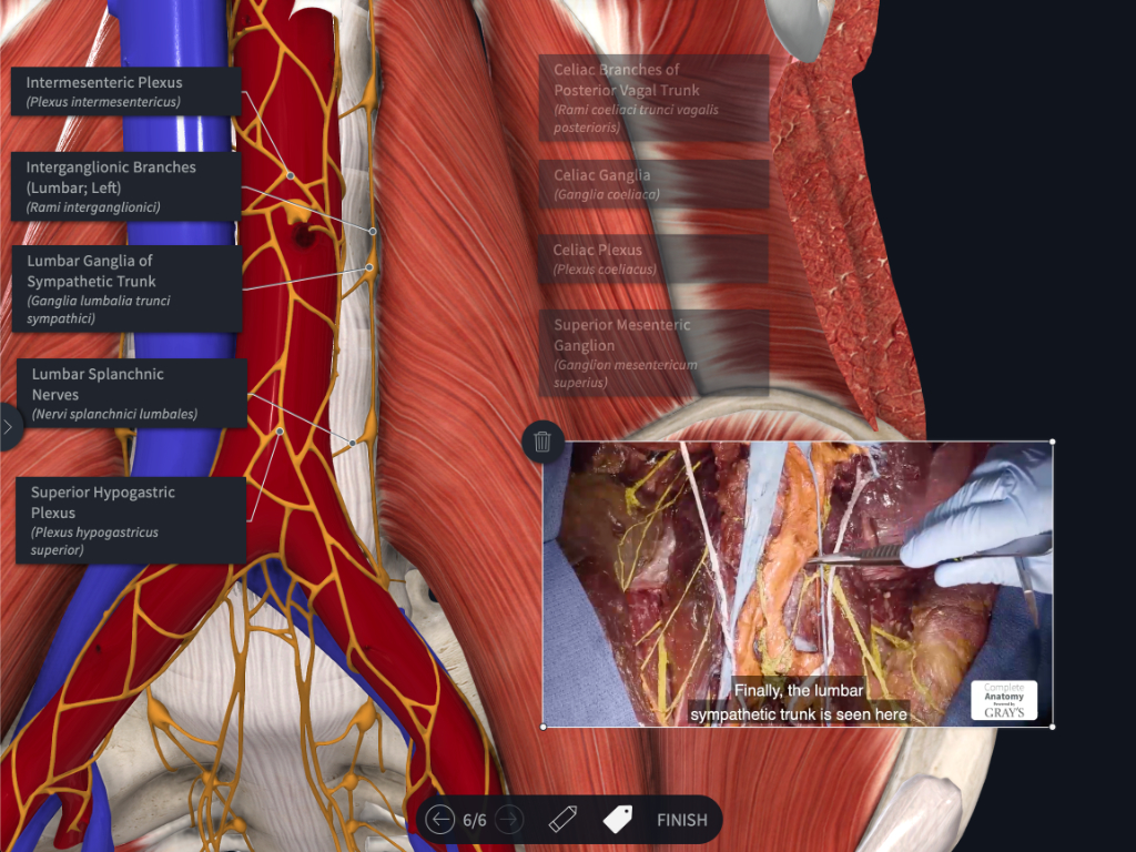
Identify the major structures which contribute to the posterior abdominal wall.

done

Understand the main blood supply, drainage, and nerve supply within the retroperitoneal space.

Author

Dr. Shane Tubbs
Dr. Shane Tubbs
Tulane University School of Medicine

R. Shane Tubbs, MS, PA-C, PhD is a native of Birmingham, Alabama, USA and a clinical anatomist, author, editor, and researcher. He is Professor of Neurosurgery, Neurology, Surgery, and Structural & Cellular Biology, Director of Surgical Anatomy at Tulane School of Medicine and Program Director of Anatomical Research in the Clinical Neuroscience Research Center at Tulane University School of Medicine, New Orleans, Louisiana. Dr. Tubbs is President-Elect of the American Association of Clinical Anatomists and serves as the Editor-in-Chief of the journal Clinical Anatomy. His h-index is 74 and in 2018, he was listed as a “hyperprolific author” in the journal Nature. He has also authored/edited over 50 books including Gray’s Anatomy Review editions 1-3, Gray’s Clinical Photographic Dissector of the Human Body editions 1 and 2, Netter’s Introduction to Clinical Procedures, the 5th through 8th editions of Netter’s Atlas of Anatomy, Nerves and Nerve Injuries volumes I and II, and Bergman’s Comprehensive Encyclopedia of Human Anatomic Variation. He is a section editor for the 41st and 42nd editions of Gray’s Anatomy. Dr. Tubbs was recently appointed Chair of the Federative International Programme on Anatomical Terminologies (FIPAT) and oversees six working groups dedicated to this topic. Under his leadership, the second edition of Terminologia Anatomica was just published.

Contents

Module 1
The Anterior Abdominal Wall
Introduction by Dr. Shane Tubbs
2 MINUTES
Lecture 1 - The anterior abdominal wall
10 MINUTES
Bony framework of abdominal region
Anterior Abdominal Wall
Layers of the anterior abdominal wall
Inguinal canal
Deep dissection of anterior abdominal wall
Dissection of peritoneal cavity
Module 2
Abdominal Cavity I
Lecture 2 - Abdominal cavity I
9 MINUTES
Position of major abdominal organs
Peritoneal attachments and ligaments of the liver
Large Intestine and Mesocolon
Blood supply and drainage of small intestine
Blood supply and drainage of midgut region of large intestine
Blood supply and drainage of hindgut region of large intestine
Relationship between superior mesenteric artery and left renal vein
Module 3
Abdominal Cavity II
Lecture 3 - Abdominal Cavity II
5 MINUTES
Extrahepatic biliary tree and assosciated relationships
Blood supply and drainage of the foregut
Examples of peritoneal supporting ligaments and potential spaces
Module 4
The Posterior Abdominal Wall and Retroperitoneal Cavity
Lecture 4 - The posterior abdominal wall and retroperitoneal cavity
10 MINUTES
Main tributaries of the inferior vena cava
Main branches of the abdominal aorta
Position of cisterna chyli
The lumbar plexus
The thoracolumbar part of autonomic division of peripheral nervous system
LECTURE 1
The Anterior Abdominal Wall

Introduction by Dr. Shane Tubbs

2 MINUTES

Lecture 1 - The anterior abdominal wall

10 MINUTES

Bony framework of abdominal region

Anterior Abdominal Wall

Layers of the anterior abdominal wall

Inguinal canal

Deep dissection of anterior abdominal wall

Dissection of peritoneal cavity

LECTURE 2
Abdominal Cavity I

Lecture 2 - Abdominal cavity I

9 MINUTES

Position of major abdominal organs

Peritoneal attachments and ligaments of the liver

Large Intestine and Mesocolon

Blood supply and drainage of small intestine

Blood supply and drainage of midgut region of large intestine

Blood supply and drainage of hindgut region of large intestine

Relationship between superior mesenteric artery and left renal vein

LECTURE 3
Abdominal Cavity II

Lecture 3 - Abdominal Cavity II

5 MINUTES

Extrahepatic biliary tree and assosciated relationships

Blood supply and drainage of the foregut

Examples of peritoneal supporting ligaments and potential spaces

LECTURE 4
The Posterior Abdominal Wall and Retroperitoneal Cavity

Lecture 4 - The posterior abdominal wall and retroperitoneal cavity

10 MINUTES

Main tributaries of the inferior vena cava

Main branches of the abdominal aorta

Position of cisterna chyli

The lumbar plexus

The thoracolumbar part of autonomic division of peripheral nervous system