About
This app is a valuable learning and reference tool for radiologists, surgeons, medical students and nurses. Anyone with an interest in human anatomy will enjoy this application. Anatomically identical scans are labeled to allow for a unique ability to compare the same anatomical landmarks on different scan types.
Navigate
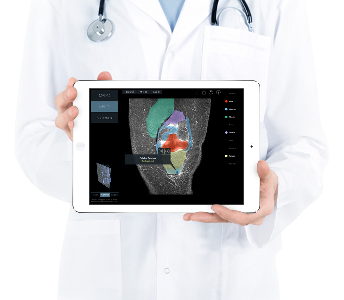
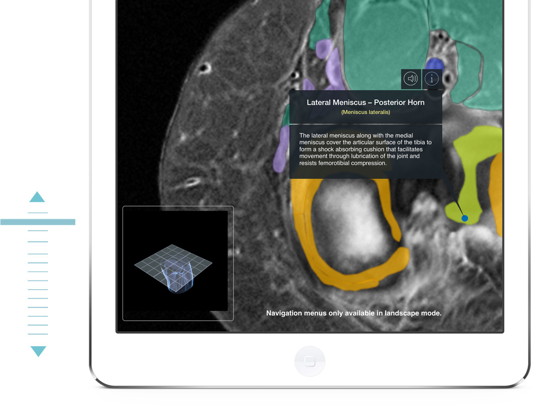
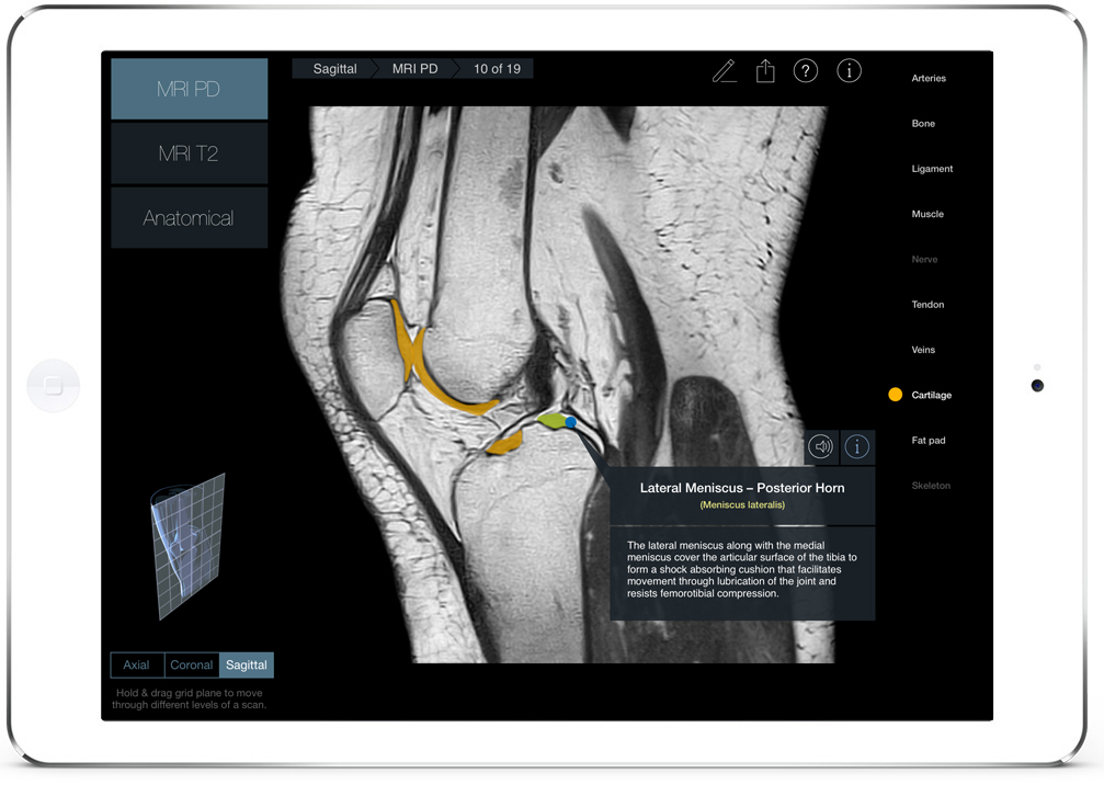
Navigate through the knee by pulling a 3D plane (Axial, Coronal or Sagittal) through a 3D knee to virtually slice through the structures. The 3D Model of the knee also allows the user to easily identify the orientation and location of the scan.
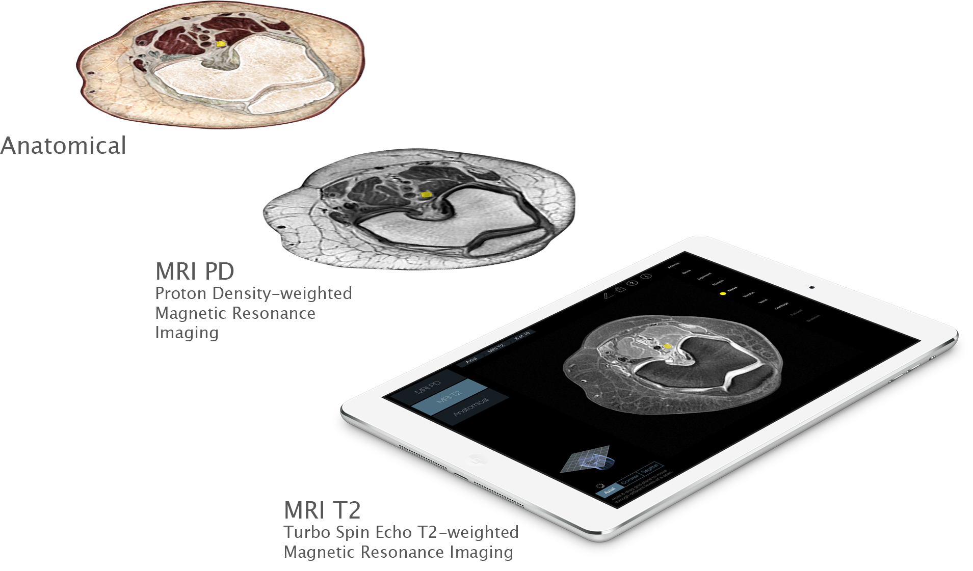
There are three planes that can be chosen, with 19 scans for each plane. These 171 images can be viewed in two different scan types, or in anatomical slices.
Compare
Radiology – Knee contains two different scan types as well as anatomical slices. These scan types are: MRI PD and MRI T2.
The scans and slices are available in all three planes: Axial, Coronal and Sagittal.
Identify
Throughout the scans are 67 individual structures that can be identified by pressing on them. Each label comes with Latin nomenclature, text description and an audio pronunciation.
Individual structures are grouped into 10 categories and each category can be turned on or off to allow for clearer identification of structures.