Complete Anatomy
Abrahams’ Clinical Human Anatomy

About this course
By Dr. Peter Abrahams
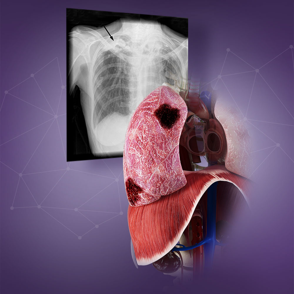
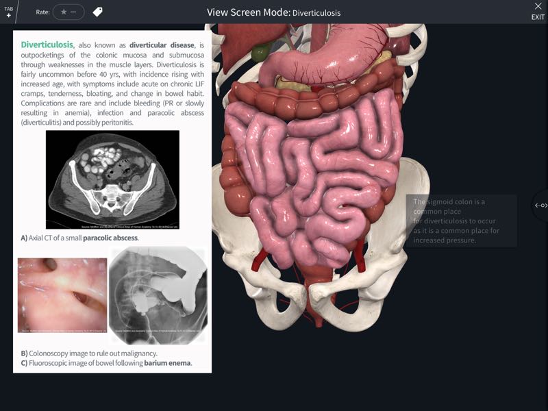
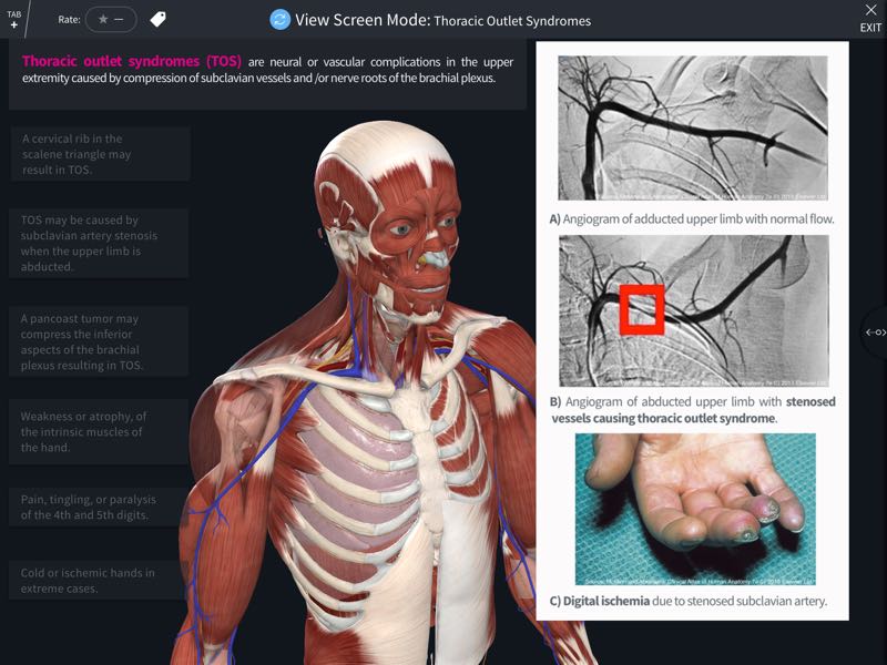
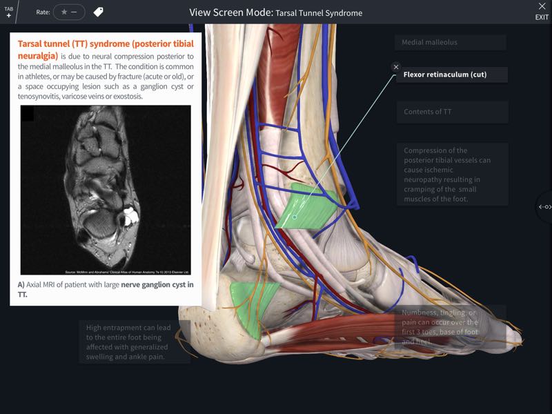
Abrahams’ Clinical Human Anatomy course includes 500 clinical images collected by the leading international anatomist Professor Peter Abrahams throughout his career. This interactive lecture series includes dynamic Screens covering essential clinical conditions which help you gain a better understanding of human anatomy by applying it to clinical practice. Correlate anatomy to clinical practice with a wealth of medical images, including X-RAY, MRI, CT, endoscopic, laparoscopic, cadaveric, ultrasound and clinical images, and explore pathologies in all regions of the body. The detailed model helps you to view the body in a more dynamic way to aid your understanding of anatomical relationships, and the excellent medical images allow you to master essential clinical conditions that every physician should know. The images utilized in the course screens correlate to the published Elsevier text McMinn & Abrahams’ Clinical Atlas of Human Anatomy.
Learning Outcomes
On completion of this course you will be able to correlate human anatomy to essential clinical conditions.


