Histology is the microscopic study of tissues and organs achieved by staining, sectioning, and viewing those sections under a microscope ?. Histology is used in scientific study, medical diagnosis, autopsy, and even in the study of fossils ?.
The four main human tissue types, epithelium, connective tissue, muscle tissue, and nervous tissue, can be viewed and distinguished with varying histological stains. There are a number of steps required before a slide can be viewed under the microscope.
Fixation
The fixation process preserves the structure of the tissue sample and prevents it from degradation by introducing chemicals that stimulate cross-linkage between proteins ?.
Dehydration
Ethanol is added to achieve dehydration of a sample ?. After the tissue sample is dehydrated, xylene can be used to remove the ethanol.
Embedding
During this phase, the tissue samples are embedded in a solid material, which allows for thin sectioning in the next stage. Paraffin wax or plastic resin are often used at this stage.
Sectioning
To section the specimen, it is placed on a precision cutting tool known as a microtome. The tissue is cut into sections of about 4-5 micrometers, which is the optimal thickness for staining.
Staining
The appropriate stain is chosen based on its unique properties. For example, Hematoxylin and Eosin are combined in H&E staining. Cell nuclei are stained blue because Hematoxylin binds to DNA, which is acidic. The cytoplasm is stained pink because Eosin binds to proteins in the cytoplasm, which are basic.
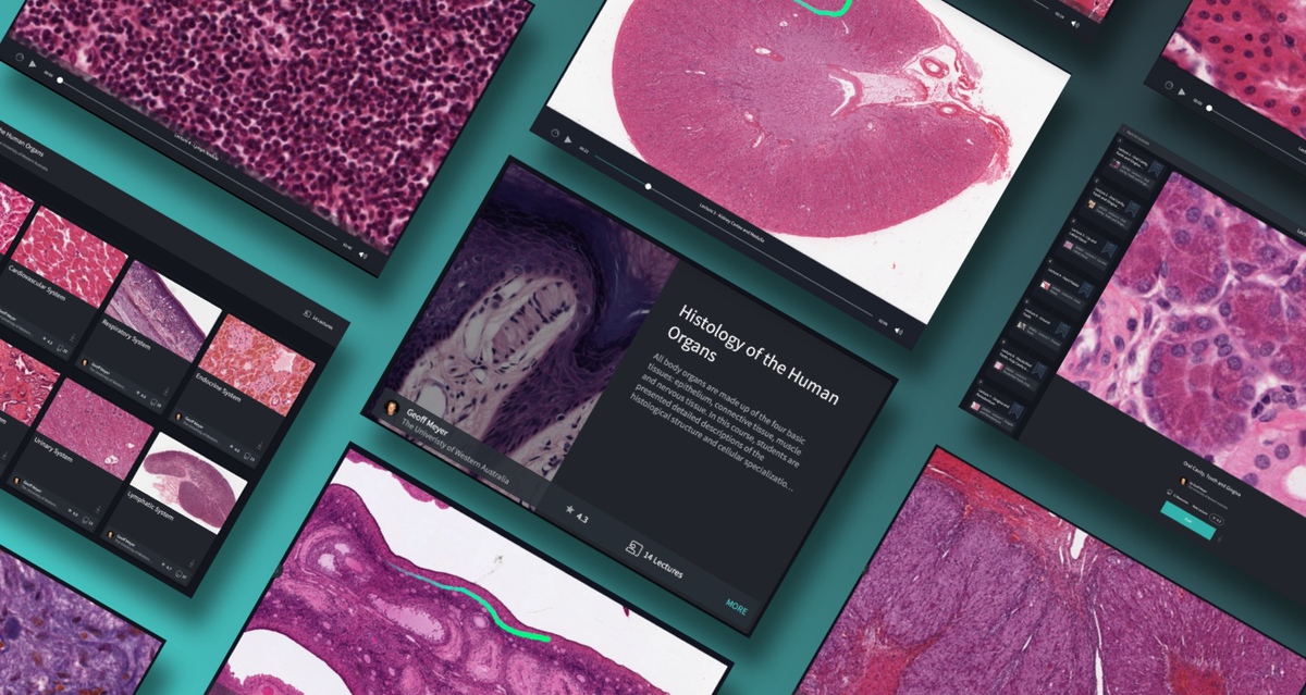
You can explore the microscopic anatomy of the human body in our Histology of the Human Organs Course, available in Complete Anatomy.