The term venipuncture (or venepuncture) is used to describe the process of entering a vein through the skin. Veni- referring to the vein, and -puncture, literally popping a hole in it ?! Veins are the the bluish vessels that run just below the skin and carry blood back to the heart. ?
There are a number of reasons why a medic might need to get access to your venous blood supply. For example, to check the number of a particular type of cells or other substances in the blood ? to deliver medicine right into the bloodstream ? or even to take a large volume when you are donating blood!
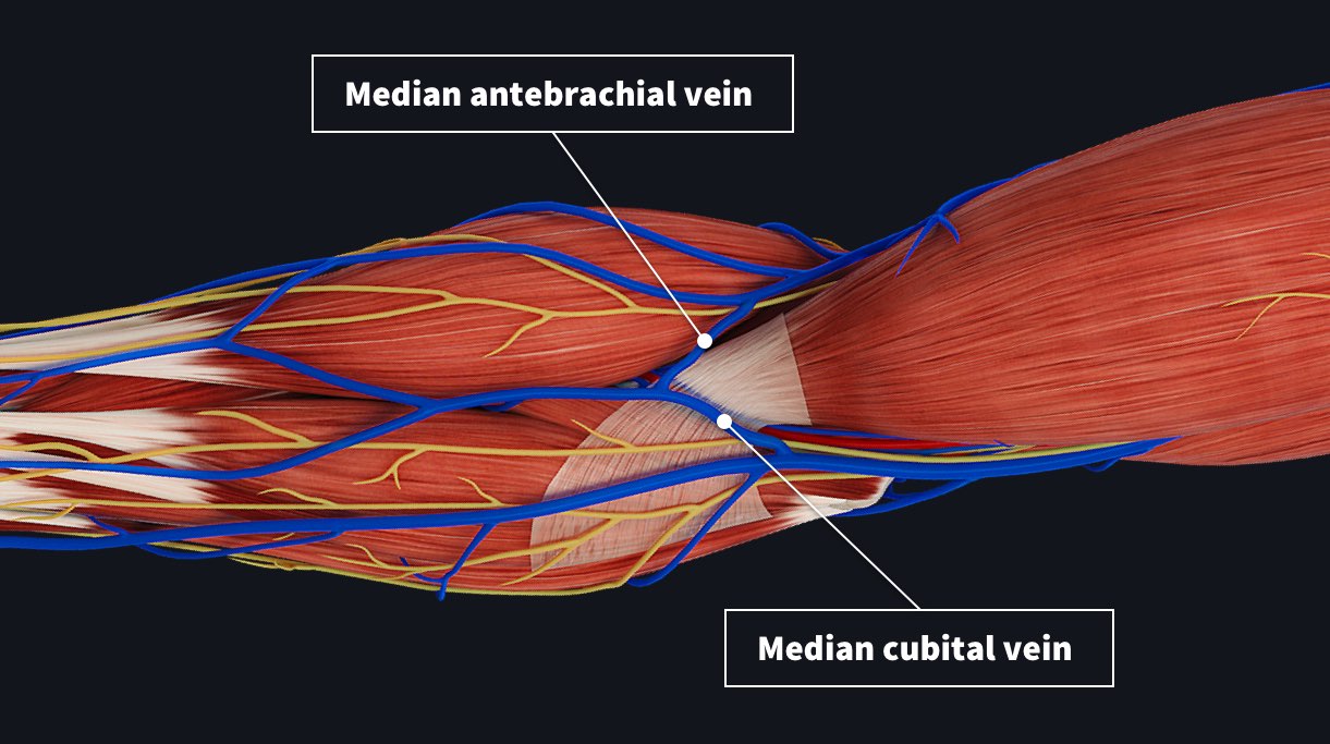
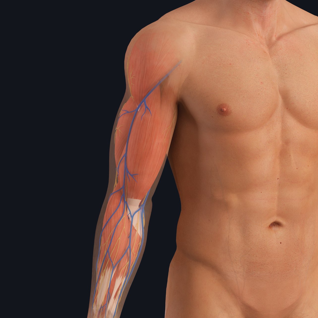
In the picture below, you can see that there is a network of blue veins just under the skin in the area in front of the elbow, the anterior cubital fossa. Blood is commonly taken from the veins that pass right across the middle of the arm: the median antebrachial vein or the median cubital vein. However, these veins present in a highly variable fashion, meaning my veins will be in a slightly different location to yours.
It’s for this reason that a clinician will usually use a tourniquet to help locate the vessels. A tourniquet is an elastic strap tied around the arm proximal to the venipuncture site. It allows blood to flow into the arm, but prevents all of it from returning to the heart because arterial blood pressure is higher than venous pressure. This causes the veins to fill up and makes them easier to see ? and palpate ?. When the clinician finds an appropriate vessel, they will stick a needle through the skin and into the vessel.
However, this isn’t always successful as the veins can wiggle away when pressure is applied to them, meaning it might take more than one attempt to find a vessel ?.
Once an adequate amount of blood has been taken, the needle is removed and pressure is applied to the site. This pressure reduces blood flow to the vessel allowing for the blood to create a small clot that will fill in the hole that the needle left in the vessel wall. If pressure is not applied, a small volume of blood will leak into the surrounding tissues causing a bruise.
Better understand the anatomy and physiology behind medical exams with Complete Anatomy. Try it for FREE today.