Skeletal muscle is one of the three main types of muscle found in the body; cardiac and smooth muscle being the other two. Skeletal muscles are under voluntary control and are responsible for moving our bodies.
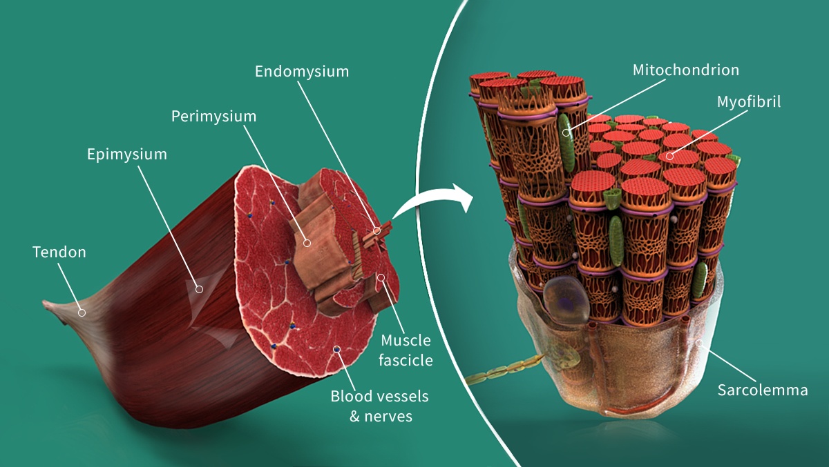
Let’s explore the anatomy of skeletal muscle starting with one of the smallest units, the myofibril. Myofibrils are contractile fibres that run parallel to each other. The myofibrils are grouped into bundles and surrounded by a plasma membrane called the sarcolemma.
A group of myofibrils makes up a unit called a muscle fibre. Muscle fibres are long, cylindrical multinucleated cells. Each muscle fibre is surrounded by a sheath of delicate connective tissue called the endomysium.
A group of muscle fibres makes up a unit called a muscle fascicle. Because the muscle fascicle is composed of multiple muscle fibres, it has greater strength and can produce more force during contraction. A connective tissue layer called the perimysium surrounds each muscle fascicle.
A group of muscle fascicles, as well as blood vessels and nerves, make up the whole skeletal muscle. The skeletal muscle is surrounded by a fibrous sheath called the epimysium. This sheath separates the muscles from surrounding structures.
The three layers of connective tissue – the endomysium, perimysium and epimysium – are composed of collagen and elastin fibres. They therefore help give skeletal muscle resistance to tensile forces. Nerves and blood vessels also travel through these layers to supply the muscle. The endomysium, perimysium and epimysium fuse together where the skeletal muscle connects to a tendon.
Check out our Skeletal Muscle and Skeletal Muscle Fibre Detailed Models in Complete Anatomy to learn more!