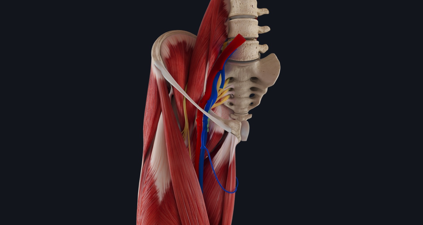
The femoral triangle?, also known as Scarpa’s triangle, is an area of much clinical importance when dealing with a femoral hernia. A femoral hernia is when part of the bowels pushes into the femoral canal. This creates a bulge within the femoral triangle. Surgical intervention? is usually needed to treat this condition.
Let’s take a closer look at the borders and contents of this space.
The femoral triangle is located anteriorly on the thigh. Three structures create its triangular shape?.
- Inguinal ligament: superior border
- Sartorius muscle: lateral border
- Adductor longus muscle: medial border and part of the floor
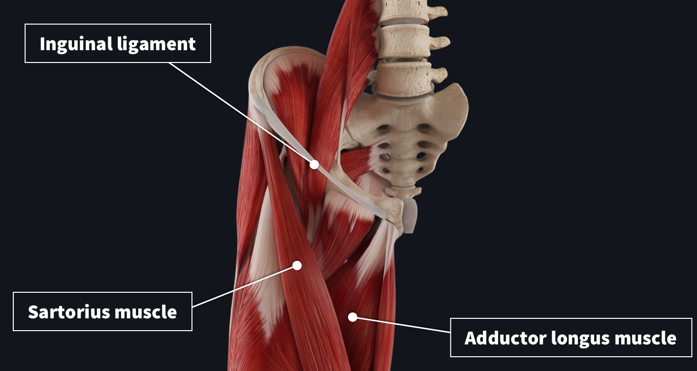
The roof consists of the facia lata, and the floor is comprised of pectineus, iliopsoas, and adductor longus.
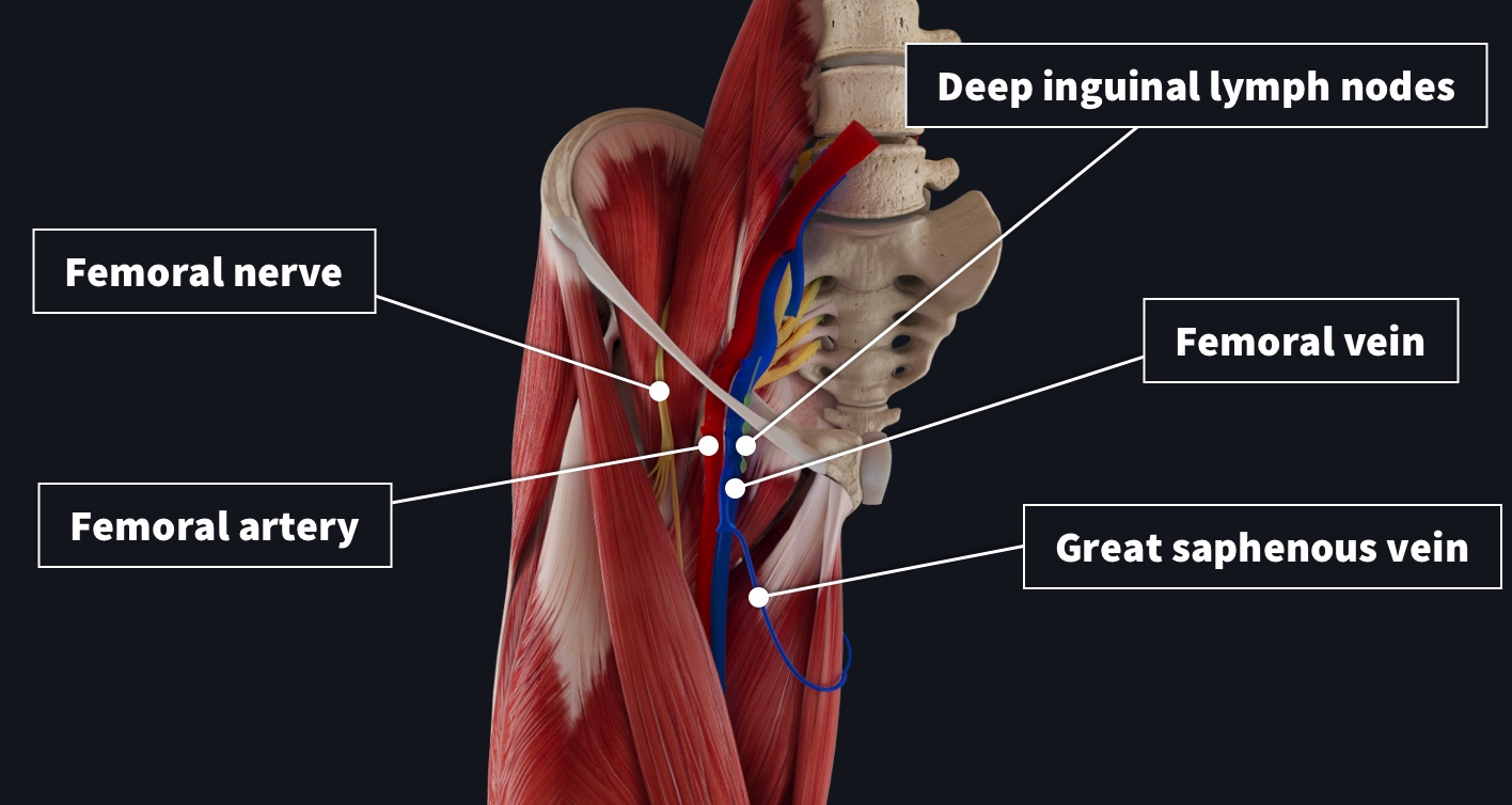
To remember the contents of the triangle, a good mnemonic to use is NAVEL.
- Nerve: femoral nerve
- Artery: femoral artery
- Vein: femoral vein (great saphenous drains into it within triangle)
- Empty space (important for vessels to distend with different levels of flow)
- Lymph canal: contains deep lymph nodes and vessels.
The femoral artery, vein, and canal are all contained within a compartment known as the femoral sheath. The sheath is made from fascia and helps protect the important vessels as they supply most of the lower limb.
If you were to palpate just inferior to where the femoral artery crosses the inguinal ligament, you can measure the femoral pulse?⚕️?⚕️.
Better understand clinical conditions such as hernias by exploring the world’s most advanced 3D anatomy platform, Complete Anatomy. Try it for FREE today.