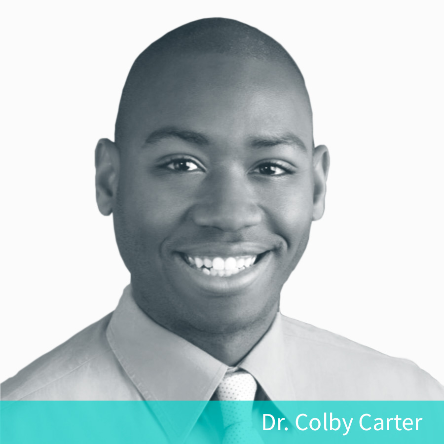
Dr. Carter started his career in medicine as a firefighter/paramedic and is a 2014 graduate of the PA program at the Medical University of South Carolina (MUSC). Due to a strong interest in Cardiothoracic surgery, he went on to be one of three in the inaugural class of Physician Assistant Surgical Residents at UF Health. He went on to study Leadership and Organizational Behavior and earned a Doctor of Health Sciences (DHSc) degree from A.T. Still University. Dr. Carter then worked as a surgical PA at UF Health and locally at St. Joseph’s Main campus.
Most recently he achieved recognition for specialty experience, skills, and knowledge through the NCCPA’s certificate of added qualifications (CAQ) specifically in Cardiovascular and Thoracic Surgery (CAQ-CVTS). In addition to teaching BLS, ACLS, and PALS, Dr. Carter currently is adjunct faculty for the PA programs at USF, Barry, and South universities as well as adjunct faculty for the EMT/Paramedic program at St. Petersburg College.
He has worked for several years in a clinical setting and more recently is involved in cardiology teaching, and is currently working with Complete Anatomy to author course content on cardiovascular surgery and neurology.
3D4Medical: How long have you been using 3D Anatomy in your educational practice? What was the turning point for you to decide to use it?
Dr. Carter: I’ve been using 3D4Medical’s applications in education since 2013 in which I used Heart Pro to educate fellow physician assistant students on structure and function of the heart. I’ve been using Complete Anatomy for about a year now. The 3D4Medical design and engineering team has continued to advance 3D anatomy to the point where I felt it was an invaluable tool for me as an educator.
“… having a 3 dimensional model that I could manipulate on my iPad was a great adjunct to explaining the pathology of their heart and how our team planned to fix it.”
3D4Medical: When you were working in a clinical setting, were you using 3D anatomy for patient education or did it come some time after that?
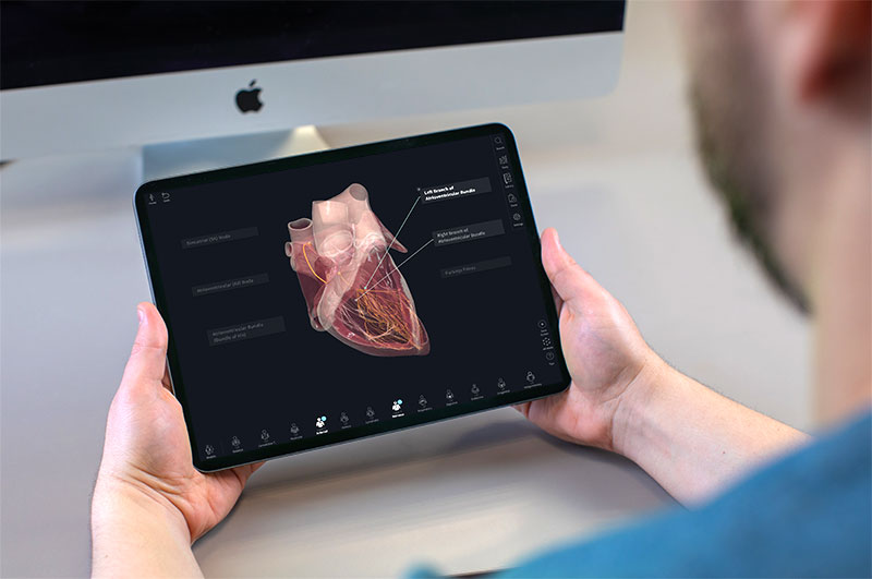
Dr. Carter: While in clinical practice as a physician assistant in Cardiothoracic surgery, one of my chief responsibilities was patient education. Specifically, I would describe in detail the surgical procedure and treatment the patient should reasonably expect. Most of my patients were 60 years and older so having a 3 dimensional model that I could manipulate on my iPad was a great adjunct to explaining the pathology of their heart and how our team planned to fix it.

3D4Medical: Now that your focus has shifted from clinical practice to cardiology teaching, what do you think are the main advantages of 3D anatomy learning for cardiology students?
Dr. Carter: The heart is a complex organ both in structure and function. How all the structures, from the chambers to valves to electrical conduction system, all interact with one another can confuse even the brightest of minds. Using 3D anatomy allows the learner, whether a patient or student, to see the structures and understand how various pathologies can result in symptomatic disease. This is done by having the ability to isolate, zoom in, and otherwise move and manipulate the model for clarity.
3D4Medical: What are the 3 main features of Complete Anatomy that you use most often when teaching?
Dr. Carter:
- I create my own Labels for structures so that when I press on them the model is made dynamic moving to a predesigned orientation. This allows me to dramatically manipulate the model for attention-grabbing visuals.
- I use the 3D pen because it allows me to highlight or demonstrate an effect with an arrow while being able to move the model and have my drawn arrow move with it.
- I teach by “flipping the classroom” in which the content of the course is delivered outside the classroom. Once we convene in the classroom, we engage in interactive activities to apply the knowledge learned from the recorded lectures. I can create custom Screens (interactive bookmarks of the model with cuts and Tools added), which allow me to thoroughly detail complex subjects such as epidemiology and etiology.
“The fact that these 3D resources are of exquisite quality, and can be used on a tablet, laptop, or smartphone anywhere at anytime, makes their widespread integration and adoption into formal medical education inevitable.”
3D4Medical: What do you think the impact of digital 3D resources will have on the future of education?
Dr. Carter: All medical or allied health students have a laptop or tablet as mandated by their institutions. Nowadays, textbooks can be downloaded in various applications, allowing a student to hold scores of medical references indexed and searchable in the palm of their hands. Digital 3D resources can be used as an adjunct or wholesale replacement for traditional medical texts such as Netter’s Anatomy or Gray’s Anatomy for the purposes of gross anatomy study. The fact that these 3D resources are of exquisite quality, and can be used on a tablet, laptop, or smartphone anywhere at anytime, makes their widespread integration and adoption into formal medical education inevitable.

3D4Medical: What are your top 3 tips for educators using Complete Anatomy?
Dr. Carter:
- Patience – there is a learning curve to the nuances of creating high-quality Screens and recordings, even for the tech savvy; but once an educator gets the hang of it, mind-blowing and engaging lectures can be developed.
- Spend extra time “getting it right” the first time when it comes to each Screen and/or recording, this saves time revising the content later.
- Create a transcript to read from when recording to make them sound more fluid and not rushed or wandering. This will also allow you to re-record more easily as medical knowledge evolves.
Dr. Carter’s Course, Cardiology / Cardiovascular Surgery, will be coming live to the platform over the next several weeks. Be the first to know about new Course releases and product updates by opting in Tips & Tricks in your email preferences.