Tears are essential for our eye health. This watery secretion contains several proteins, such as lysozymes and immunoglobulin A, which protect the surface of the eye from infection, while maintaining the hydration of the epithelial surface of the eye.
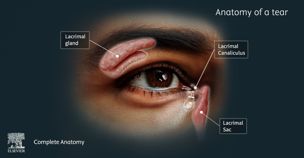
The lacrimal gland is a lobulated exocrine gland that is responsible for producing tears. The gland is divided into two portions: the orbital part and the palpebrae part, which are separated by the levator palpebrae superioris muscle. The lacrimal gland is located in the superior lateral corner of the orbit, extending out into the upper eyelid.
The gland itself contains the secretory unit, the acini, that discharge their aqueous product into a central lumen. Numerous lumens converge on an intercalated duct that opens into the conjunctival sac and is how tears are expelled from the gland. Myoepithelium surrounds the acini and ducts, and these provide mechanical force to expel tears. There are as many as twelve ducts for each gland.
Each eye contains a superior and inferior lacrimal canaliculus. The lacrimal canaliculi are responsible for draining tears from the medial angle of the eye into the nasolacrimal drainage system. The canaliculi transport the tears to the lacrimal sac, and then onto the nasolacrimal duct which directs the tears to the inferior meatus of the nasal cavity.
During crying, tear secretion increases and the output of the nasolacrimal duct increases. This causes the tears and mucus to flow out the nostrils, resulting in a runny nose. If the capacity to drain tears is overwhelmed, they spill over the margin of the lower eyelid and onto the cheeks.
Check out Complete Anatomy to learn more about the anatomy of a tear!